Untangling the loops of intestinal obstruction

 Intestinal Obstruction

One difficult and potentially dangerous condition that stands out in the complex field of gastrointestinal health is an intestinal obstruction. By discussing the definition, causes, symptoms, diagnosis, and significance of prompt intervention for successful management, this blog seeks to clarify the intricacies surrounding intestinal obstruction.
Intestinal obstruction


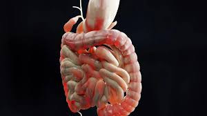
A condition known as bowel obstruction occurs when the bowels become narrowed and unable to function normally. The constriction could be brought on by a malignant tumour or colon inflammation. Food particles cannot pass through the bowel (tube) if it becomes blocked, which could result in symptoms.

 Intestinal Obstruction Causes:


  • After abdominal surgery, scar tissue may form, which may cause the intestines to adhere to one another and obstruct flow.
  • An obstruction could result from a section of the intestine poking through a weak spot in the abdominal wall.
  • The normal flow may be impeded by benign or malignant growths in the surrounding tissues or within the intestine.
  • It is possible for the colon to twist on itself, obstructing the blood flow and causing a blockage.
  • Conditions such as diverticulitis or Crohn's disease can cause inflammation, which can then result in blockage.

Symptoms of intestinal obstruction

It is essential to recognise the symptoms of intestinal blockage in order to get medical help as soon as possible. Typical signs and symptoms consist of:


  •  Lower abdominal pain, either intermittent or constant, can cause cramping.
  • Vomiting may result from an inability to pass gas or stool.
  • Abdominal distension and swelling brought on by trapped fluid and gas.
  • inability to pass stool or lack of bowel movements.
  • a discernible decrease in gas passage.

Diagnosis & Treatment

Intestinal blockage diagnosis is made by combining medical history, physical examination, and diagnostic imaging tests (endoscopy, CT scan, or X-ray). Prompt medical intervention is imperative upon identification in order to avert complications like ischemia and bowel perforation.


To remove the fluids and gas from the patient's intestine, it is imperative to keep them off of food and to use a nasogastric tube.

The patient will receive intravenous fluids to treat their dehydration and maintain normal electrolyte levels.

Patients may occasionally receive medication to treat underlying infections that are the cause of their problems, and if other treatments are unsuccessful, surgery may be performed.


To remove a bowel obstruction, surgery may be necessary. This could include stoma formation, adhesiolysis (adhesion release), intestinal resection, or a bypass surgery. In order to suture (join) the two ends of the bowel together, surgeons may remove the damaged section. They will make an abdominal incision to remove the bowel's end, and a stoma bag will collect your stool if the two ends cannot be joined.


Metallic tubes, also known as stents, called self-expanding metal stents (SEMS) are used to hold the colon open when a tumour obstructs it so that stools can pass through.

Recovery Phase

Intestinal obstruction recovery frequently entails a mix of lifestyle and medication changes. Recurrence must be avoided by post-treatment, dietary changes, and routine check-ups with medical professionals.


Take away point

Being aware of the symptoms and risk factors can encourage people to seek care as soon as possible, increasing the likelihood that their treatment will be successful and reducing the likelihood of complications such as intestinal perforation or aspiration of bowel contents into stomach.For a comprehensive assessment and individualised care, speak with a medical professional if you believe you may be experiencing symptoms of intestinal obstruction.


Transient Ischemic Attacks Do Put Your Life At Danger??

What are Transient Ischemic Attacks??


Transient Ischemic Attacks (TIAs) are important warning indicators in the complex field of neurological health that should never be disregarded. TIAs, sometimes called "mini-strokes," are brief interruptions in blood supply to the brain that present a critical chance for intervention and the avoidance of more serious strokes. This blog post will go over the particulars of transient ischaemic attacks, including their causes, symptoms, and the importance of acting quickly to avoid long-term harm.

Brief Introduction

 When there is a brief interruption in the blood supply to a portion of the brain, transient ischemic attacks take place. The effects of a TIA are transient, typically lasting a few minutes to several hours, in contrast to a full-blown stroke. TIAs, however, should not be taken lightly as they frequently signal a vascular issue that requires medical attention.

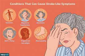
Transient ischemic attacks (TIAs) and strokes share many of the same symptoms.. A TIA is referred to as a mini-stroke by some. In contrast to stroke symptoms, which are typically more permanent, a TIA's symptoms disappear entirely within 24 hours. A small blood clot in a brain blood vessel is the most frequent cause.


It's critical that you act quickly to see if the symptoms resolve on their own. If your symptoms have improved rapidly and totally, you should make an appointment with your doctor right away for additional testing and care.

The symptoms of a transient ischemic attack (TIA) are brief and result from a brief loss of blood supply to a portion of the brain. While the symptoms are similar to those of a stroke, they are transient and disappear quickly. This indicates that you've fully recovered and are back to normal. (A diminished blood and oxygen supply to a portion of the body is referred to as an ischemic state.)

Causes of TIA 


A tiny blood clot that gets lodged in a brain artery or small blood vessel is typically the cause of a transient ischemic attack (TIA). As a result, part of the brain is oxygen-starved and the blood flow is blocked. Only a few minutes are spent without oxygen in the affected area of the brain before it fully recovers. This is because the blood clot either disintegrates rapidly or the blood vessels in the vicinity can dilate.

These clots can result from a number of conditions, including carotid artery disease, atrial fibrillation (an irregular heartbeat), and atherosclerosis (narrowing of the arteries). They can also originate from the heart or major blood vessels.

On a patch of atheroma in a major neck artery, a small blood clot frequently forms. Taking blood to the brain are the major arteries in the neck, the carotid and vertebral arteries. It is possible for the small blood clot to separate from the atheroma or for a small portion of the blood clot to break off. After that, the bloodstream carries this on its way to the brain. The clot travels until it gets stuck as the arteries gradually get smaller. It clogs the artery and cuts off the blood supply when it gets stuck.


If a small blood clot becomes stuck, it is likely to disintegrate rapidly. As a result, a TIA causes no long-term brain damage, and symptoms quickly disappear.

Blood clot Formation  

A blood clot may form if platelets stick to an atheroma patch (plaque).  Blood platelets are tiny particles that help clot blood in the event that a vein or artery is severed. Atheroma plaques are like the accumulation of scale inside your kettle; they are fatty lumps that form inside the lining of arteries.  Periodically, a clot is formed when platelets adhere to an atheroma plaque inside an artery.


Symptoms 

A TIA's symptoms are momentary, or transient. They often reach their zenith in less than a minute after emerging abruptly. Though symptoms can last anywhere from two to fifteen minutes, they usually subside within an hour. At times, symptoms can persist for a full day. The affected brain region determines the symptoms that appear. Various bodily parts are under the control of distinct brain regions. Consequently, a few or all of the following symptoms could be present:

  •     A hand, arm, or leg's weakness or clumsiness.
  •     communication difficulties.
  •     swallowing difficulties.
  •     A portion of the body that feels numb or pins and needles.
  •     momentary blindness or double vision.


Investigation Done In TIA

Typically, the following tests are performed:

  •   Blood tests.  Your doctor may first perform a blood sugar test to rule out low blood sugar, which can mimic the symptoms of a transient internal affair (TIA).
  •  Additionally, a blood test will be performed to rule out diabetes and high cholesterol, two conditions that increase the risk of TIA and stroke. 


  • Additionally, tests will be performed to ensure that you do not have kidney issues or iron deficiency anaemia. These are the most significant blood tests, though there are occasionally others performed. Generally speaking, CT brain scanning is not advised unless your specialist believes it could detect something other than TIA. 


  • This is to check whether atheroma has caused severe narrowing of one of these arteries. Similar to fatty lumps, atheroma plaques form inside the arterial lining. A risk factor for TIA and stroke is carotid artery narrowing, or stenosis. In the event that one or both of your carotid arteries are over 50% clogged, you might receive an urgent referral for unclogging surgery. 
  • CT scan can reveal which areas of the brain were damaged, whether there was a brain bleed, or whether there was another issue (like a brain tumour) that resulted in your symptoms.
  • An ECG to look for irregular cardiac rhythms, such as atrial fibrillation, which is an irregular heartbeat. Your doctor's office may handle this.


  • Measurement of blood pressure. While your blood pressure won't technically be tested, it's still important to have it checked. Treatment for high blood pressure is necessary because it increases the risk of TIA and stroke.

Treatment 


In order to address the underlying risk factors, treatment frequently focuses on taking blood thinners to prevent blood clot formation, cholesterol, and blood pressure medications. Important elements of managing TIAs include making lifestyle changes like eating a heart-healthy diet, exercising frequently, and giving up smoking.


Healthy Living Following a TIA

Individuals are frequently counselled to alter their lifestyles in order to lower their chance of having another incident after suffering a transient episode. Maintaining a healthy and resilient future is facilitated by frequent check-ins with medical professionals, taking prescription drugs as directed, and continuously monitoring risk factors. 


Individuals are frequently counselled to alter their lifestyles in order to lower their chance of having another incident after suffering a transient episode. Maintaining a healthy and resilient future is facilitated by frequent check-ins with medical professionals, taking prescription drugs as directed, and continuously monitoring risk factors.


Knowing the Mysteries about Erythema Nodosum

Introduction 

A skin condition known as erythema nodosum (EN), which affects the layer of fat beneath the skin, is classified as panniculitis. Even though the term "condition" sounds complicated, it's important to understand for people who might experience its symptoms or who want to learn more about their dermatological health.


The hallmark of erythema nodosum is the development of painful, tender, reddish-purple nodules or lumps, usually on the shins. These nodules often develop symmetrically on both legs and range in size. The most common location is the shins, but it can also affect the thighs, forearms, and trunk.

It is assumed that erythema nodosum is a hypersensitivity reaction. It frequently appears as a dermatological symptom of an infectious or other illness.The incidence of erythema nodosum differs between different countries. The peak incidence occurs between the ages of 20 and 40, though it can happen at any age. It is roughly five times more common in adult women than in adult men. In kids, the frequency is the same for boys and girls.


Symptoms & Morphologhy of Rash

An excruciating rash is followed by fever, aching, and arthralgia during the eruptive phase of erythema nodosum.

Lesions start off as sensitive, red nodules. The borders have a diameter of 2–6 cm and are ill-defined.


The initial week of erythema nodosum is characterised by hard, tense, and painful lesions. In the second week, they may become fluctuant, much like an abscess, but they do not suppurate or ulcerate. In most cases, lesions disappear after two weeks, but occasionally they reappear after three or six.

Weeks may pass while you still have aching legs and swollen ankles. Initially, they are a vivid crimson, but after a week or two, they take on a blue or purple tint, and they even turn yellow, akin to a healing bruise, before eventually going away.


 More than half of patients experience joint pains, which usually start two to four weeks prior to the eruptive phase. The joints may have effusions and are tender, swollen, and red. There could be stiffness in the morning. Most frequently affected areas are the wrists, ankles, and knees. While synovitis goes away in a few weeks, stiffness and pain in the joints can linger for up to six months. The joint has not undergone any detrimental alterations, and the synovial fluid lacks cells.

Underlying causes

As a reaction to an underlying trigger, EN is a reactive condition rather than a stand-alone disease:


  • Inflammatory bowel disease, Behçet's disease, pregnancy, drug reactions, streptococcal infection, and primary TB are the most frequent causes.
  • Erythema nodosum can also be caused by sarcoid.
  • Erythema nodosum can manifest clinically in leprosy, but the lesions' histological appearance is distinct.
  • EN can be brought on by bacterial and viral infections, such as fungal infections, TB, or streptococcal throat infections.A higher risk of developing EN is linked to conditions like autoimmune such as sarcoidosis and inflammatory bowel disease.
  • It typically appears in the second half of pregnancy. It may happen when using oral contraceptives and is likely to recur in subsequent pregnancies.
  • Some cases  have been linked to HIV, hepatitis B, hepatitis C, and Epstein-Barr virus.

Investigations

 
   Send an ESR and FBC. Regardless of the cause, ESR is frequently very high, and CRP might have a greater role.

Despite the possibility of a negative result even in cases of streptococcal illness, a throat swab is a suitable test for streptococcus.
The anti-streptococcal O (ASO) titre could be more beneficial, even though a normal titre does not rule out infection. Perhaps more valuable is a rising titre.


Tests for Y. enterocolitica, Salmonella spp., and Campylobacter spp. in the stool and blood cultures may provide results; however, since most labs will not process formed stool for infection, this is usually only worthwhile if the patient has diarrhoea.
Angiotensin-converting enzyme (ACE) and calcium levels are frequently elevated in sarcoidosis.
In cases of sarcoidosis, a chest X-ray may reveal evidence of pulmonary tuberculosis, unilateral or asymmetrical adenopathy in cases of malignancy, or bilateral hilar lymphadenopathy (BHL).


To rule out coccidioidomycosis and tuberculosis, intradermal skin tests might be necessary.
When there is uncertainty about the diagnosis, an excisional biopsy may be useful.


A complete medical history, physical examination, and occasionally additional tests, like blood tests, chest X-rays, or skin biopsies, are part of the diagnosis process in order to determine the underlying cause.

Treatment 

Taking care of the underlying cause is the main objective of EN treatment. Most cases are self-limiting and require only symptomatic relief.It may be necessary to prescribe antibiotics or antifungal drugs if EN is secondary to an infection. In autoimmune disease cases, taking care of the underlying illness becomes essential.


    Symptomatic Relief: Nonsteroidal anti-inflammatory medicines (NSAIDs) or corticosteroids can be used to treat the pain and inflammation brought on by EN.
    Supportive Measures: Applying cold compresses, elevating the afflicted limbs, and getting rest can all help reduce symptoms.

Many potential treatments for persistent cases of erythema nodosum are listed in the literature; these include oral steroids, tetracyclines, macrolides, potassium iodide, and biologic drugs. Nevertheless, it would seem prudent to consult secondary care at this point to ensure that no underlying diagnosis is being overlooked.Erythema nodosum typically goes away in six weeks, but it can take longer in some cases, particularly if it is idiopathic or the underlying cause is still present.


Take away Point

Try to find the main causes of the rash if you start to notice one on your body so that it can be better managed. However, it will typically settle down on its own, so there's no need to worry.


Have You Heard About Familial Obesity?

Breaking the Cycle to Promote Future Health 


Millions of people around the world suffer from obesity, a complicated and multidimensional health problem. Familial obesity refers to obesity that runs in families, implying a genetic susceptibility to this condition. The purpose of this blog is to examine the different facets of familial obesity, including its causes, effects, and methods for ending the cycle in order to encourage a healthier future.


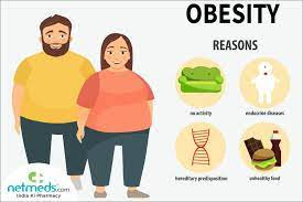
There are some factors contributing towards this:

Genetic Predisposition 

A genetic component is frequently present in familial obesity, as some people may inherit a propensity to gain weight. Although certain genes are linked to obesity, environmental factors and lifestyle choices also have a big impact on the disease's development. 


Your own life style

Familial obesity is influenced not only by genetics but also by shared family environments and lifestyles. Weight management can be greatly influenced by eating habits, levels of physical activity, and family health-related culture as a whole.


Comedian James Corden recently conducted the topic of obesity in a monologue, highlighting the hereditary component of the condition. He emphasised that people who are genetically predisposed to obesity are struggling against both their biology and social stigma, and he called for a change in emphasis from appearance to better health. This viewpoint casts doubt on the idea that people should be held responsible for their weight and emphasises how crucial it is to comprehend the intricate interactions between heredity, environment, and societal perceptions of obesity.


Effects of Obesity On You

 Numerous health hazards, such as an elevated risk of cardiovascular disease (CVD), hypertension, dyslipidemia, thromboembolic events, and type 2 diabetes, are linked to familial obesity.It becomes essential to break the cycle in order to reduce these risks.


Familial obesity can cause psychological problems for affected individuals, including low self-esteem, problems with body image, and a higher risk of mental health disorders like depression. For overall well-being, it is essential to address both the psychological and physical components.


 Studies have indicated that there is a strong correlation between obesity and family history, with children of overweight parents having an 80% chance of becoming overweight themselves.  Furthermore, obesity can affect fertility and the health of future generations and is linked to a higher risk of dying young. It's crucial to understand that, although genetics plays a big part in familial obesity, other factors like lifestyle decisions and the family environment also play a part. Individuals and families may minimise the influence of genetic predisposition on general health by promoting healthy habits and raising awareness of the health risks linked to familial obesity.


What is Your Share? 

If you're lost, try not to worry and just be part of this journey in the following ways:

  •  In order to break the cycle of familial obesity, it is important to promote healthy habits within the family. Promoting a healthy diet, frequent exercise, and positive body image attitudes are all part of this.


  •  It is imperative to increase knowledge regarding the genetic and environmental components that lead to obesity. Families with educated members are better able to make decisions and lead healthier lives.
  • Consulting nutritionists, dietitians, and fitness specialists among other healthcare professionals can offer individualised advice. Families can work with them to create attainable and long-lasting weight management strategies.
  •  Taking care of one's emotional health is essential to ending the family obesity cycle. A healthier mindset is facilitated by encouraging open communication, creating a supportive environment, and fostering a positive relationship with food.


  • Acknowledging minor triumphs along the journey is crucial. It can inspire people and families to stick with their health goals when they recognise and value even the smallest improvements.Facilitating this journey for yourself and those who accompany you can be transformative.


Take away Point

To live a healthier lifestyle, keep in mind that it takes time, and every step in the right direction leads to a better, healthier future.Are you ready to undertake a significant life change?


Know Why You Got Pancreatitis and How You can Manage it?

Overview

 An inflammation of the pancreas, which is in charge of generating digestive enzymes and controlling blood sugar, is the hallmark of pancreatitis.  Behind the stomach is an organ. Pancreatitis is a disease that develops when the pancreas becomes inflamed.Pancreatitis comes in two primary forms: acute and chronic. Acute pancreatitis is typically transient and may result from gallstones or alcohol consumption, whereas chronic pancreatitis is a chronic illness frequently associated with heavy alcohol use and smoking.


Causes of Pancreatitis 

There are some possible causes of pancreatitis:


  • One of the most frequent causes of pancreatitis is gallstones. Inflammation can result from these tiny, hardened deposits obstructing the ducts that empty the pancreas. 
  • Pancreatitis is also largely caused by excessive alcohol consumption. Drinking alcohol can harm the pancreas over time, increasing the possibility of inflammation.


  • It can be brought on by abdominal injuries or physical trauma. It could result from surgical procedures, falls, or accidents.
  • The pancreas may also become inflamed due to bacterial or viral infections. The mumps or specific parasite infections can cause pancreatitis.
  • In certain instances, pancreatitis may be influenced by genetic factors. People who have a family history of the illness might be more vulnerable. 


Presentation of This Condition

 The signs and symptoms of acute or chronic pancreatitis can differ. Both types' primary symptom is excruciating upper abdominal pain that can radiate to the back. Pain can start slowly or come on suddenly in people with acute pancreatitis, while severe pain that gets worse after eating can be felt continuously in people with chronic pancreatitis. Jaundice, or yellowing of the skin and eyes, is another common symptom along with fever, fast heartbeat, nausea, and vomiting.  This might be the outcome of the pancreas inflammation causing disturbances in the digestive system. Intentional weight loss and severe dehydration can result from severe pancreatitis cases.


 An elevated body temperature and an accelerated heart rate can result from pancreatitis. These signs show how the body is reacting to inflammation. Jaundice, which is defined by skin and eye yellowing. This happens when the neighbouring bile ducts are impacted by an inflammatory pancreas.

 Treatment Options 

  • For the pancreas to heal, rest is necessary. In order to achieve this, stay as still as possible and abstain from food and liquids.
  •  An important part of treating pancreatitis is managing pain. Medication for pain and alterations in lifestyle, like abstaining from alcohol and eating a low-fat diet, can help control symptoms.Acetaminophen or ibuprofen are two examples of painkillers that can help with pain relief. Stronger painkillers, like morphine, might be required in extreme situations.


  • Effective treatment of pancreatitis requires determining and treating its underlying cause. This could entail alcohol dependency management programmes or surgery to remove gallstones.
  • Individuals suffering from pancreatitis might need nutritional assistance, particularly if their pancreas isn't operating at its best. Dietary changes, enzyme supplements, or, in extreme situations, intravenous nutrition may be necessary for this.
  •  


  • Hospitalisation may be necessary for severe pancreatitis cases. This enables medical staff to closely monitor the patient, administer intravenous fluids, and handle any complications.

Living with Pancreatitis

Chronic pancreatitis is a chronic illness that needs to be managed continuously. Although the pancreas cannot heal itself and is permanently damaged, there are steps that can be taken to manage the disease and lower the risk of complications. 


This covers dietary modifications, lifestyle adjustments, and routine check-ups with the doctor4. In summary, pancreatitis is a dangerous illness that needs to be managed with medical care and lifestyle changes. For those who are impacted by this illness, it is essential to comprehend the signs, causes, and available treatments. For a precise diagnosis and suitable treatment, it's critical to seek medical attention as soon as you or someone you know exhibits pancreatitis symptoms. 


Time to seek Medical help

If you experience persistent or unexpected stomach pain, schedule a visit with your physician. If you can't sit still due to extreme pain, get medical attention right away and find a more comfortable position.